Hi! I’ve finished up the third week of my internship at the Frazier Rehab Institute. Unfortunately, about halfway through the week I was quarantined, but I got to experience so many different things before then! I assisted with the setup of a patient in a machine that was new to me, and then got to observe how that machine collected the data that was being transmitted by the patient’s neurological reaction to stimuli (electric currents). Additionally, I helped set up and witnessed what happens during an MMR, which involved direct stimulation to the patients back where the spinal cord is located. I also had an amazing time at the Louisville Zoo with my host family before getting quarantined, which I am so grateful for. I got to see so many animals!
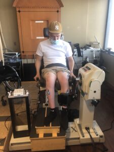
In the photo below you may notice small blue boxes with wires attached to the participant’s legs. These sensors detect the muscle contractions that take place when those muscles are stimulated. This stimulation was introduced into the patient’s legs and lower abdomen, and would help maximize the efforts made by the participant to move their legs.
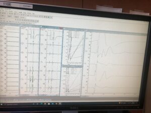
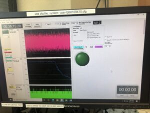
In order to properly record the information produced by the muscle contractions of the paralyzed individual, the data gathered by the blue sensors was collected and displayed on the computer screens shown below. This way the torque (blue line in second photo below), measurable force produced by the participant’s muscle contractions, could be interpreted in relation to different variables like when the electric currents of the stimulation were introduced into the patient’s legs (pink line). The graphs in the first photo presented below represent the different areas of the leg that were being stimulated in relation to the sensors that were placed over those muscles.
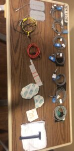
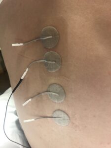
As mentioned previously, I observed an MMR take place for the first time. The first photo displayed below demonstrates how the materials required during an MMR would be prepared. This includes different electrodes, adhesives (tape), alcohol pads, and a razor, which is used to prep the location of the participant’s skin that will have some of these electrodes. In addition to the patient’s legs receiving stimulation, there were also electrodes placed on the back (as shown in the second photo) where their spinal cord is located to transmit signals directly to the central nervous system. By the way, the central nervous system includes the brain and spinal cord, while the peripheral nervous system is a general term used to categorize all other neurons and has different, more specific components like the somatic nervous system.
This week I also got to visit the Louisville Zoo for the first time and got to learn about so many new animals. I was taught by my host family’s kids that groups of penguins in the water as shown below are known as a raft, but when they’re on land they are identified as a waddle. I had such an amazing time at my internship and with my host family this week, and I can’t wait for next week!


There are no comments published yet.