It is baffling to think that I am currently a third of the way through my six-week internship; time really does fly. Spending time in the lab over the last two weeks has given me a thorough taste of the different research projects that are in progress, and of all of the behind-the-scenes work that is essential to the proper functioning of the study (which has turned out to be a lot more than I originally expected). Despite the occasional tedium of the process, taking part in the lab continues to be extremely fascinating and enjoyable.
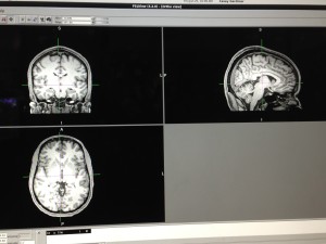
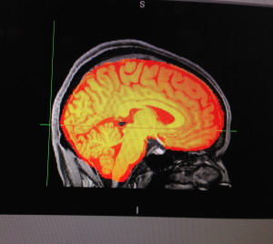
This last week, in addition to witnessing neurocognitive tests and another fMRI, I was able to help QC (‘quality control’) an fMRI scan. In order to do so, we used a program called ‘FSL’ to scan through the final images created from the fMRI, and looked for any abnormalities that could indicate computer malfunctions, movement, or tumors. In addition, we also brought up computer-made graphs of people’s movement during the fMRI. If the participant moved over two millimeters during the scan, the images were trashed.

fMRI images of the human brain during a QC (quality control) session
Quick fact: do you know why the human brain looks the way it does (shriveled up and walnut-like)? No? Well prepare to be mind-blown. The brain essentially folds in on itself in order to maximize its surface area. Picture a balloon deflating and tucking in on itself in order to form a furrow-like pattern. This particular appearance gives the human brain enhanced processing power, thus enabling humans to act as we do rather than like other animals in the animal kingdom. This is one of the many facts that have mind-boggled me over the last few weeks.

An fMRI image shaded orange to detect abnormalities
Overall, I am unbelievably excited to have this opportunity; I am looking forward seeing what the next few weeks have in store.

There are no comments published yet.